
- Anatomical Deficit and Limitations of Classical Keratoplasty
- PB-001 Technology and Bioprinting Architecture from Precise Bio
- Structural Features of the Printed Implant
- Clinical Trials at Rambam Health Care Campus
- Monitoring Results and Regeneration Assessment in 2026
- Financial Performance and Investment Potential of the Sector
Anatomical Deficit and Limitations of Classical Keratoplasty
Corneal transplantation is one of the most frequent operations in the field of vision restoration, but global ophthalmology faces a critical shortage of donor material. Statistical data indicate that for one available donor, there are more than 130 patients who need replacement of damaged or damaged corneal tissue. In addition to the acute shortage, classic keratoplasty is accompanied by the risk of immune rejection of the transplant, because the recipient’s body can recognize foreign proteins even despite the low level of vascularization of the corneal membrane. The use of fully synthetic polymer implants also does not solve the problem in the long term, as artificial materials often provoke chronic inflammatory processes, glaucoma, or extrusion of the endoprosthesis.
The development of regenerative medicine allowed moving from the use of inorganic materials to the creation of bioengineered matrices. The initial stages of research were based on purified collagen of animal origin, which served as a scaffold for subsequent colonization by patient cells. However, the real technological shift was the transition to a full-fledged three-dimensional bioprinting, where living human cells functioning in specially developed hydrogels are used as the main building material.
PB-001 Technology and Bioprinting Architecture from Precise Bio
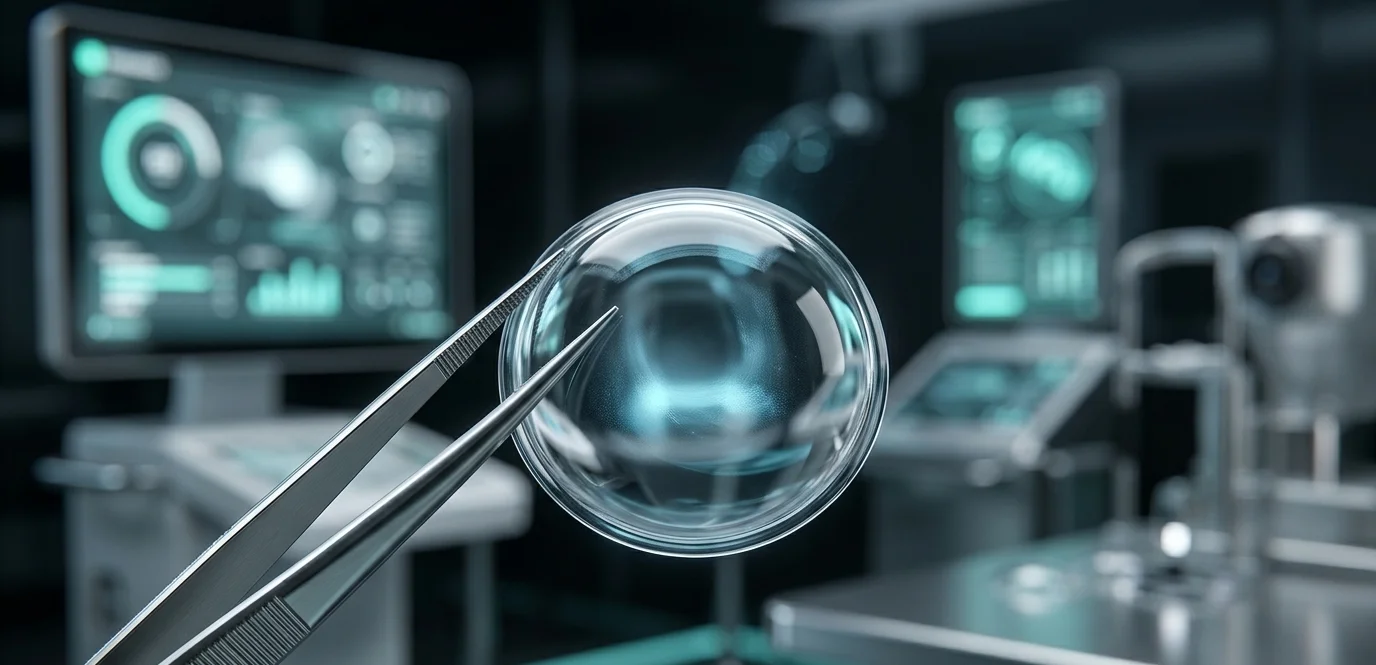
The Israeli biotechnology company Precise Bio changed the approach to the creation of eye tissues by developing its own engineering platform for printing living structures. The product, which received the index PB-001, is the first cornea in the world completely printed on a 3D bioprinter using cultured human cells. The process is based on a unique technology of spatial positioning of cells, which allows reproducing the complex multilayer architecture of a natural organ.
Structural Features of the Printed Implant
The natural human cornea consists of five main layers, each of which performs a clear optical and barrier function. The technological process of PB-001 bioprinting is focused on accurate modeling of the stroma and endothelial layer. To achieve the necessary optical and mechanical properties, the following components are used
- Cell Material: Highly purified human keratocytes, which are responsible for the synthesis of extracellular matrix and maintenance of tissue transparency.
- Bioink: A specialized hydrogel based on natural polymers, which ensures the viability of cells during printing and preserves the given geometry until full polymerization.
- Endothelial Layer: A single layer of cells on the inner surface of the implant that regulates the hydration level of the cornea, preventing its swelling.
The main advantage of the platform is scalability. Using cell material from only one donor cornea unsuitable for direct transplantation, the laboratory is able to grow and print up to 300 high-precision PB-001 implants. This completely negates the problem of organ shortage and significantly reduces the cost of preparation for surgery.
Clinical Trials at Rambam Health Care Campus
The transition of technology from laboratory conditions to real medical practice took place on the basis of the Rambam Medical Center in Haifa under the leadership of Professor Michael Mimouni. The first-ever implantation of a bioprinted cornea into a human was performed as part of Phase 1 clinical trials, the main purpose of which is to assess the safety, biocompatibility, and optical stability of the implant.
The operation was performed on a patient with end-stage corneal disease, when residual vision was minimal and standard treatment methods did not yield results. Surgical intervention was performed according to the standard protocol of penetrating keratoplasty, which confirms the integration of the new material into the existing medical infrastructure. The bioprinted tissue demonstrated high elasticity and resistance to surgical suturing, which was previously a problem for pure collagen analogues.
Monitoring Results and Regeneration Assessment in 2026
During 2026, researchers conducted regular monitoring of the patient’s condition after implantation. The key criteria for success are the absence of a rejection reaction, the gradual engraftment of the implant cells into the surrounding tissues of the recipient, and the restoration of transparency of the optical medium of the eye.
It was recorded that the bioprinted structure PB-001 did not cause an acute immune response. Due to the precise geometry and optical uniformity, surface roughness is minimized, which allowed achieving a stable focusing ability. The engraftment process is accompanied by a gradual replacement of the hydrogel matrix with the patient’s own collagen, which indicates the launch of natural regenerative processes inside the eyeball.
Financial Performance and Investment Potential of the Sector
The development of technologies for three-dimensional bioprinting of eye tissues is supported by large investment funds and grants. Precise Bio attracted significant funding from strategic partners, including major ophthalmic corporations in the United States and Europe. The total volume of investments in the development of the PB-001 line and related printed tissues (including the retina) exceeded 50 000 000 USD. According to market analysts, the commercialization of this technology after the completion of all phases of clinical trials will reduce the average cost of vision restoration surgery by 40%, making high-tech care more accessible to patients worldwide.




0 Comments